Fraud Blocker Skip to main content

Mouse Mat > Fine Art Storehouse > Science Inspired Art

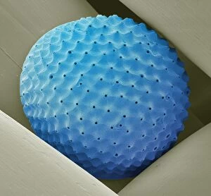
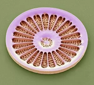
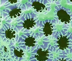
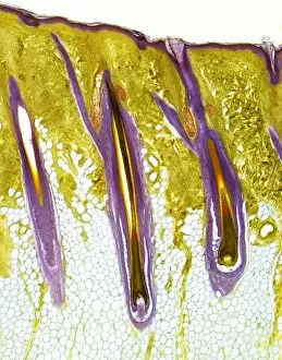
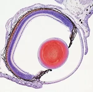
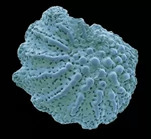
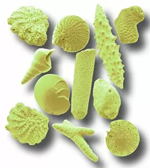
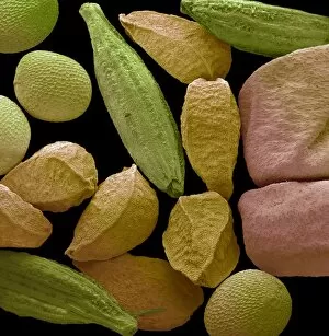
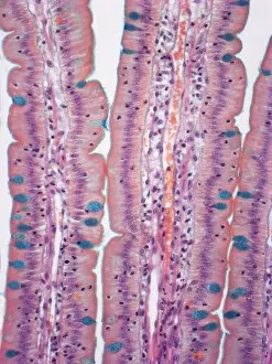
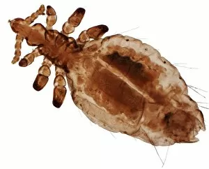
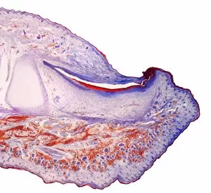
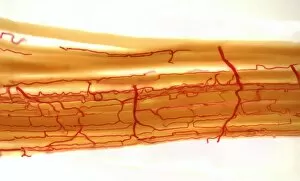
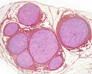
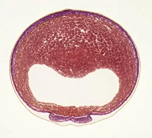
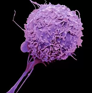
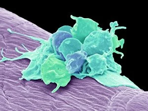
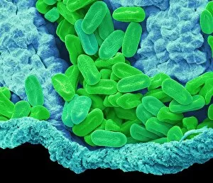
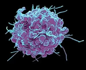
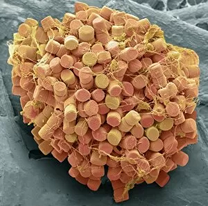
Science Inspired Art Mouse Mat Collection (#9)

Showcase your love of Science with prints from the fields of biology, physics, mathematics, space, medicine and more

1,282 Mouse Mats

We are proud to offer this selection in partnership with Fine Art Storehouse

Background imageScience Inspired Art Mouse Mat Collection: Radiolarian, SEM

Radiolarian, SEM
Radiolarian. Coloured scanning electron micrograph (SEM) of a radiolarian. Radiolarians are amoeboid protozoa that produce intricate mineral skeletons

Background imageScience Inspired Art Mouse Mat Collection: Diatom, SEM

Diatom, SEM
Diatom. Coloured scanning electron micrograph (SEM) of a single diatom (Arachnoidiscus sp.). Diatoms may be extremely abundant in both freshwater

Background imageScience Inspired Art Mouse Mat Collection: Diatom detail, SEM

Diatom detail, SEM
Diatom. Coloured scanning electron micrograph (SEM) of detail of the silica wall of a diatom. Diatoms may be extremely abundant in both freshwater

Background imageScience Inspired Art Mouse Mat Collection: Human skin showing hair follicles, LM

Human skin showing hair follicles, LM
Hairy skin. Light micrograph of a thick section of human skin, showing three central hair follicles. The outer layer of the skin, the epidermis, is the thin, purple band, supported by the deeper

Background imageScience Inspired Art Mouse Mat Collection: Frog eye, light micrograph

Frog eye, light micrograph
Frog eye. Light micrograph of a section through the eye of a frog. Most amphibians, such as frogs and salamanders, have color vision

Background imageScience Inspired Art Mouse Mat Collection: Foraminiferan microfossil, SEM

Foraminiferan microfossil, SEM
Foraminiferan. Coloured scanning electron micrograph (SEM) of a foraminiferan microfossil from maldives beach sand. Microfossils are roughly 0.05 to 2mm in size

Background imageScience Inspired Art Mouse Mat Collection: Microfossils, SEM

Microfossils, SEM
Sand microfossils. Coloured scanning electron micrograph (SEM) of microfossils from maldives beach sand. Microfossils are roughly 0.05 to 2mm in size

Background imageScience Inspired Art Mouse Mat Collection: Panch phoran, SEM

Panch phoran, SEM
Panch phoran. Coloured scanning electron micrograph (SEM) of panch phoran or indian five spice. It is a spice blend commonly used in Eastern India and Bangladesh and consists of the following seeds

Background imageScience Inspired Art Mouse Mat Collection: Sea salt, SEM

Sea salt, SEM
Sea salt. Coloured scanning electron micrograph (SEM) of sea salt, showing its crystalline structure. Sea salt consists mainly of sodium chloride (NaCl), but unlike pure table salt

Background imageScience Inspired Art Mouse Mat Collection: Diatoms and radiolaria, SEM

Diatoms and radiolaria, SEM
Diatoms and radiolaria. Coloured scanning electron micrograph (SEM) of a circular arrangement of various diatoms and radiolaria. Diatoms are planktonic unicellular algae

Background imageScience Inspired Art Mouse Mat Collection: Virus particles and bacteria, SEM

Virus particles and bacteria, SEM
Vaccinia virus particles and bacteria. Coloured scanning electron micrograph (SEM) of vaccinia virus particles (green). Unlike most viruses, vaccinia replicates in the cells cytoplasm

Background imageScience Inspired Art Mouse Mat Collection: Flower bud, LM

Flower bud, LM
Flower bud. Light microscope image (LM) of a section of a flower bud. The small yellow pollen grains are visible in the anthers, the male part of the flower

Background imageScience Inspired Art Mouse Mat Collection: Obelia medusa, LM

Obelia medusa, LM
Obelia hydrozoan medusa. Light micrograph (LM) of a medusa (young polyp) from the Obelia geniculata hydroid. The circular shallow semi-bell has solid tentacles on the rim

Background imageScience Inspired Art Mouse Mat Collection: Developing nail, LM

Developing nail, LM
Developing nail. Light micrograph (LM) of longitudinal section through a fetal finger tip to show the developing nail. The large area of green-yellow nail bed epithelium is tipped by the developing

Background imageScience Inspired Art Mouse Mat Collection: Onion root tip, LM

Onion root tip, LM
Mitosis. Light micrograph (LM) of a transverse section of onion (Allium cepa) root tip to show cells undergoing mitosis (nuclear division). Magnification: x100 when printed at 10 centimetres wide

Background imageScience Inspired Art Mouse Mat Collection: Liverwort spore capsule, LM

Liverwort spore capsule, LM
Liverwort spore capsule. Light micrograph (LM). Longitudinal section through the thallus and sporangium of a liverwort (Pellia epiphylla)

Background imageScience Inspired Art Mouse Mat Collection: Small intestine, LM

Small intestine, LM
Small intestine. Light micrograph (LM) of a section through the finger-like projections (villi) of the duodenum, the uppermost part of the small intestine

Background imageScience Inspired Art Mouse Mat Collection: Human head louse, LM

Human head louse, LM
Human head louse, Light microraph (LM) 0f Pediculus humanus capitis. P. humanus is divided into two subspecies; the head louse P. humanus capitis and the body louse P. humanus corporis

Background imageScience Inspired Art Mouse Mat Collection: Thyroid, LM

Thyroid, LM
Thyroid gland. Light micrograph (LM) of a thyroid gland showing the follicles. The follicles are lined by a single layer of cuboidal epithelial cells (blue)

Background imageScience Inspired Art Mouse Mat Collection: Fingertip, LM

Fingertip, LM
Fingertip. Light micrograph (LM) of a section through the fingertip. The nail (orange) is at top center, with the nail root below. The nail bed is dark purple and is continous with the epithelium

Background imageScience Inspired Art Mouse Mat Collection: Blood supply to muscles, LM

Blood supply to muscles, LM
Blood supply to muscles. Light micrograph (LM) showing blood supply to muscle fibers. The muscle fibers (yellow) have been teased apart to reveal the capillary bed (red)

Background imageScience Inspired Art Mouse Mat Collection: Fallopia tube, LM

Fallopia tube, LM
Fallopian tube. Light micrograph (LM).The fallopian tube, or oviduct, conveys the egg from the ovary to the uterus. Ciliated columnar epithelium is yellow

Background imageScience Inspired Art Mouse Mat Collection: Mushroom gill, LM

Mushroom gill, LM
Mushroom gills. High power light micrograph (LM) of a section through the gills of a mushroom, Agaricus sp. (formerly Psalliota sp.)

Background imageScience Inspired Art Mouse Mat Collection: Spinal cord, LM

Spinal cord, LM
Spinal cord. Light micrograph (LM) of a cross-section through the human spinal cord in the lumbar region. The spinal cord consists of a butterfly-shaped core (dark blue) known as grey matter

Background imageScience Inspired Art Mouse Mat Collection: Trachel epithelium, LM

Trachel epithelium, LM
Trachea epithelium. Light micrograph (LM) of a vertical section through the pseudostratified columnar epithelium from the trachea

Background imageScience Inspired Art Mouse Mat Collection: Female flea, LM

Female flea, LM
Human flea. Light micrograph (LM) of a female human flea (Pulex irratans). Fleas are wingless and flattened from side to side, which makes them difficult to dislodge in hair

Background imageScience Inspired Art Mouse Mat Collection: Nasal sinuses, LM

Nasal sinuses, LM
Nasal sinuses. Light micrograph (LM) of the nasal sinuses ( lined by cyan epithelium ) and the supporting cartilages (green). Bone tissue is identified by the blue bone marrow

Background imageScience Inspired Art Mouse Mat Collection: Xylem, LM

Xylem, LM
Xylem tissue. Light micrograph (LM) of a section through sunflower(helianthus annuus) tissue showing spiral tracheids, a type of xylem

Background imageScience Inspired Art Mouse Mat Collection: Taste buds, LM

Taste buds, LM
Taste buds. Coloured light micrograph of a section through the tongue, showing taste buds (round, yellow). The taste buds are within papillae (projections) located on the surface of the tongue

Background imageScience Inspired Art Mouse Mat Collection: Nerve fibres, LM

Nerve fibres, LM
Nerve fibres. Light micrograph (LM) of a transverse section through a bundle (fascicle) of nerve fibres. Within each fascicle are many myelinated nerve fibres

Background imageScience Inspired Art Mouse Mat Collection: Developing frog egg, LM

Developing frog egg, LM
Developing frog egg. Light micrograph of a transverse section through a developing egg laid by a common frog (Rana temporaria). The egg has reached the early neurula stage

Background imageScience Inspired Art Mouse Mat Collection: Macrophage, SEM

Macrophage, SEM
Macrophage. Coloured scanning electron micrograph (SEM) of a macrophage white blood cell. Macrophages are cells of the bodys immune system

Background imageScience Inspired Art Mouse Mat Collection: Activated platelets, SEM

Activated platelets, SEM
Activated platelets. Coloured scanning electron micrograph (SEM) of activated platelets attached to surgical gauze. Platelets are tiny blood cells that help the body form clots to stop bleeding

Background imageScience Inspired Art Mouse Mat Collection: Streptococcus mutans, SEM

Streptococcus mutans, SEM
Streptococcus mutans. Coloured scanning electron micrograph (SEM). S. mutans is a coccoid shaped, Gram-positive, anaerobic bacteria that is part of the normal bacteria flora of the mouth

Background imageScience Inspired Art Mouse Mat Collection: Plaque-forming bacteria, SEM

Plaque-forming bacteria, SEM
Plaque-forming bacteria, coloured scanning electron micrograph (SEM). Plaque consists of a film of bacteria embedded in a glycoprotein matrix

Background imageScience Inspired Art Mouse Mat Collection: Fallopian tube, SEM

Fallopian tube, SEM
Fallopian tube. Coloured scanning electron micrograph (SEM) of the surface of a human fallopian tube. Fallopian tubes are ducts that lead from the ovaries to the uterus

Background imageScience Inspired Art Mouse Mat Collection: C elegans, SEM

C elegans, SEM
Caenorhabditis elegans worm, coloured scanning electron micrograph (SEM). C. elegans is a soil-dwelling hermaphrodite nematode worm and one of the most studied animals in biological

Background imageScience Inspired Art Mouse Mat Collection: Soil bacteria, SEM

Soil bacteria, SEM
Soil bacteria. Coloured scanning electron micrograph (SEM). Bacteria in the soil are directly tied to nutrient recycling especially carbon, nitrogen, phosphorus and sulfur

Background imageScience Inspired Art Mouse Mat Collection: Resting T lymphocytes

Resting T lymphocytes. Coloured scanning electron micrograph (SEM) of resting T lymphocytes from a human blood sample. T lymphocytes, or T cells

Background imageScience Inspired Art Mouse Mat Collection: Bird bone tissue, SEM

Bird bone tissue, SEM
Bird bone tissue. Coloured scanning electron micrograph (SEM) of cancellous (spongy) bone from a starlings (Sturnus vulgaris) skull

Background imageScience Inspired Art Mouse Mat Collection: Kefir bacteria, SEM

Kefir bacteria, SEM
Kefir bacteria. Scanning electron micrograph (SEM) of Lactococcus bacteria from kefir, a fermented milk beverage containing beneficial yeast as well as probiotic bacteria

Background imageScience Inspired Art Mouse Mat Collection: Bacteria found on mobile phone, SEM

Bacteria found on mobile phone, SEM
Bacteria found on mobile phone. Coloured scanning electron micrograph (SEM) of bacteria cultured from a mobile phone. Tests have revealed the average handset carries 18 times more potentially harmful

Background imageScience Inspired Art Mouse Mat Collection: Nematodes, SEM

Nematodes, SEM
Phasmarhabditis hermaphrodita. Coloured scanning electron micrograph (SEM) of Phasmarhabditis hermaphrodita. Phasmarhabditis hermaphrodita is a microscopic nematode in the family Rhabditidae

Background imageScience Inspired Art Mouse Mat Collection: Tendon, SEM

Tendon, SEM
Tendon, coloured scanning electron micrograph (SEM), showing bundles of collagen fibres. The parallel alignment of the fibres make tendons inelastic but flexible. Tendons attach muscle to bone

Background imageScience Inspired Art Mouse Mat Collection: Brochosomes, SEM

Brochosomes, SEM
Brochosomes, coloured scanning electron micrograph (SEM). Brochosomes are intricately structured microscopic granules secreted by leafhoppers

Background imageScience Inspired Art Mouse Mat Collection: Verbena pollen, SEM

Verbena pollen, SEM
Verbena pollen. Coloured scanning electron micrograph (SEM) of pollen grains from verbena bonariensis. Verbena bonariensis is a tall, slender-stemmed perennial

Background imageScience Inspired Art Mouse Mat Collection: Apoptosis, SEM

Apoptosis, SEM
Apoptosis. Coloured scanning electron micrograph (SEM) of a 293T cell in the early stages of programmed cell death, or apoptosis. Apoptosis occurs when a cell becomes old or damaged

Background imageScience Inspired Art Mouse Mat Collection: Diatoms, SEM

Diatoms, SEM
Diatoms. Coloured scanning electron micrograph (SEM) of fresh water centric diatom frustules (skeleton), Diatoms are a type of algae (Chromophyta, Bacillariophyceae)


All Professionally Made to Order for Quick Shipping


-

Why Choose Us?

We are a leading provider of Art Prints and Photo Gifts since 2003, working in partnership with a range of Sporting Clubs, Charities, Museums and Picture Libraries. A large share of profits from any of their images will go directly towards supporting that charity or club. Our archive of images is carefully curated to bring you a wide range of subjects, including landscapes, wildlife, architecture, and more. We ship from our partner labs in the UK, USA, EU (Netherlands) and Australia.
+

How do I place an order?

Ordering is quick & easy - Just follow 5 Simple Steps:
  1. Find your image: Use our search box or browse our online photo Collections to find the image you want.

  2. Choose your print format: Select your desired product and add it to your cart.

  3. Enter your details: If you're a returning customer, simply enter your email address and password, and we'll fill in your billing and shipping address details. All personal details are held securely and are fully GDPR compliant. As standard, we remove all Personally Identifiable Information after 12 months.

  4. Pay for your purchase: We use state-of-the-art security for online shopping and do not have access to your card details.

  5. Sit back and relax: We'll email you confirmation of your order and when it's dispatched. Registered customers can also track orders in the 'My Account' area.

+

How do I pay for an order?

You can pay for your order with most of the major credit and debit cards, or PayPal. For added security, major financial institutions process payment details separately and securely on our behalf. We do not have access to your online payment card details. Online payments are preferred however we do take phone orders. For UK customers only we accept cheques issued against a UK bank.
+

Is my data safe?

We take data security very seriously. We do not have access to your full card details and all payments are requested over a fully secure connection. Additionally, we fully comply with current European and GDPR legislation, and automatically remove all personal data after at most 12 months (unless you have an account with us and have signed in recently). We also have a strict opt-in policy and would never sell your personal details. Your data is only used to fulfill your order promptly and efficiently.
+

Is wall art ready to hang?

For quick and easy installation all wall art, including framed prints, canvas prints and metal prints are supplied with a ready to hang solution on the back. Generally, saw tooth hangers are applied as they allow wall art to hang flush against the wall. The serrated edge of the hanger prevents the frame from shifting or tilting when hung.
+

Are the photo prints fade resistant?

Yes, we use archival quality photo paper photographic paper for vivid reproduction Prints are an accurate representations of the original artwork, which is preserved for artistic character and authenticity. We guarantee they match previews shown on our web site