Fraud Blocker Skip to main content

Home > Fine Art Storehouse > Science Inspired Art > The Human Brain

The Human Brain Collection (#4)

Illustrations of the Human Brain, the command center for the human nervous system and one of the largest and most complex organs in the human body. It is made up of more than 100 billion nerves that communicate in trillions of connections called synapses

210 Items

We are proud to offer this selection in partnership with Fine Art Storehouse

Background imageThe Human Brain Collection: Digital illustration of human brain

Digital illustration of human brain

Background imageThe Human Brain Collection: Digital illustration of head in profile showing pituitary gland in brain highlighted in blue

Digital illustration of head in profile showing pituitary gland in brain highlighted in blue

Background imageThe Human Brain Collection: Digital illustration of reward pathway in human brain

Digital illustration of reward pathway in human brain

Background imageThe Human Brain Collection: Digital illustration of human brain associated with full awareness

Digital illustration of human brain associated with full awareness

Background imageThe Human Brain Collection: Digital illustration of areas of information highlighted in human brain

Digital illustration of areas of information highlighted in human brain

Background imageThe Human Brain Collection: Digital illustration of human brain with orbitofrontal cortex and amygdala highlighted in green

Digital illustration of human brain with orbitofrontal cortex and amygdala highlighted in green

Background imageThe Human Brain Collection: Digital illustration of head in profile showing areas of brain used for processing emotion

Digital illustration of head in profile showing areas of brain used for processing emotion

Background imageThe Human Brain Collection: Digital illustration of head in profile showing brain

Digital illustration of head in profile showing brain

Background imageThe Human Brain Collection: Digital illustration of head in profile showing mirror neurons in human brain highlighted in

Digital illustration of head in profile showing mirror neurons in human brain highlighted in purple, pink and green

Background imageThe Human Brain Collection: Digital illustration of head in profile showing area of mirror neuron in brain highlighted in orange

Digital illustration of head in profile showing area of mirror neuron in brain highlighted in orange

Background imageThe Human Brain Collection: Digital illustration of head in profile showing direction of sensory signals from visual cortex to

Digital illustration of head in profile showing direction of sensory signals from visual cortex to hippocampus in brain

Background imageThe Human Brain Collection: Digital illustration of monism and dualism in human brain

Digital illustration of monism and dualism in human brain

Background imageThe Human Brain Collection: Digital illustration of prefrontal cortex of human brain highlighted in green

Digital illustration of prefrontal cortex of human brain highlighted in green

Background imageThe Human Brain Collection: Digital illustration of various areas of cortex in human brain receiving input from sense organs

Digital illustration of various areas of cortex in human brain receiving input from sense organs

Background imageThe Human Brain Collection: Digital illustration of crucial parts of highlighted in human brain

Digital illustration of crucial parts of highlighted in human brain

Background imageThe Human Brain Collection: Digital illustration of frontal lobe and parietal lobe areas (orange) in left hemispheres (blue)

Digital illustration of frontal lobe and parietal lobe areas (orange) in left hemispheres (blue), and pathway of data from parietal lobe to frontal lobe (green) in human brain

Background imageThe Human Brain Collection: Digital illustration of lateral prefrontal cortex involved in decision making highlighted in green

Digital illustration of lateral prefrontal cortex involved in decision making highlighted in green in human brain

Background imageThe Human Brain Collection: Digital illustration of premotor cortex involved in decision making highlighted in pink in human

Digital illustration of premotor cortex involved in decision making highlighted in pink in human brain

Background imageThe Human Brain Collection: Digital illustration of human head in profile showing brain

Digital illustration of human head in profile showing brain

Background imageThe Human Brain Collection: Digital illustration of right superior temporal sulcas, and anterior cingulate cortex highlighted

Digital illustration of right superior temporal sulcas, and anterior cingulate cortex highlighted in red and grey in human brain

Background imageThe Human Brain Collection: Digital illustration of anterior insular, anterior cingulate cortex

Digital illustration of anterior insular, anterior cingulate cortex, and ventromedial prefrontal cortex highlighted in human brain

Background imageThe Human Brain Collection: Digital illustration of premotor cortex involved in decision making highlighted in pink in human

Digital illustration of premotor cortex involved in decision making highlighted in pink in human brain

Background imageThe Human Brain Collection: Digital illustration of head in profile showing skull, brain, and spine

Digital illustration of head in profile showing skull, brain, and spine

Background imageThe Human Brain Collection: Digital illustration of head in profile showing brain and spine

Digital illustration of head in profile showing brain and spine

Background imageThe Human Brain Collection: Digital illustration of head in profile showing brain and spine

Digital illustration of head in profile showing brain and spine

Background imageThe Human Brain Collection: Digital illustration of head in profile showing hippocampus and amygdala (green)

Digital illustration of head in profile showing hippocampus and amygdala (green), and auditory area (red) in brain

Background imageThe Human Brain Collection: Digital illustration of head in profile showing skull, brain and spine

Digital illustration of head in profile showing skull, brain and spine

Background imageThe Human Brain Collection: Digital illustration of head in profile showing auditory cortex (red), and hippocampus (green)

Digital illustration of head in profile showing auditory cortex (red), and hippocampus (green) and direction of signals in brain

Background imageThe Human Brain Collection: Digital illustration of unconscious and conscious pathways in human brain passing through thalamus

Digital illustration of unconscious and conscious pathways in human brain passing through thalamus ending in parietal lobe of cortex

Background imageThe Human Brain Collection: Digital illustration of direction of dopamine flow, nucleus accumbens, basal ganglia

Digital illustration of direction of dopamine flow, nucleus accumbens, basal ganglia and ventral tegmental area highlighted in human brain

Background imageThe Human Brain Collection: Black and white digital illustration of cingulate cortex area of human brain

Black and white digital illustration of cingulate cortex area of human brain

Background imageThe Human Brain Collection: Digital illustration of amygdala in human brain

Digital illustration of amygdala in human brain

Background imageThe Human Brain Collection: Digital illustration of human brain with spine highlighted in pink

Digital illustration of human brain with spine highlighted in pink

Background imageThe Human Brain Collection: Digital illustration of head in profile showing brain

Digital illustration of head in profile showing brain

Background imageThe Human Brain Collection: Digital illustration of human brain with primary auditory cortex highlighted in orange and red

Digital illustration of human brain with primary auditory cortex highlighted in orange and red

Background imageThe Human Brain Collection: Digital illustration of head in profile showing memory areas of brain

Digital illustration of head in profile showing memory areas of brain

Background imageThe Human Brain Collection: Digital illustration of head in profile showing amygdala, auditory cortex, wernickes area

Digital illustration of head in profile showing amygdala, auditory cortex, wernickes area, and anterior temporal lobe in human brain

Background imageThe Human Brain Collection: Digital illustration of head in profile showing bundle of nerve fibres connecti ng Brocas area

Digital illustration of head in profile showing bundle of nerve fibres connecti ng Brocas area and Wernickes area in human brain

Background imageThe Human Brain Collection: Digital illustration of areas associated with memory in human brain

Digital illustration of areas associated with memory in human brain

Background imageThe Human Brain Collection: Digital illustration of human brain

Digital illustration of human brain

Background imageThe Human Brain Collection: Digital illustration of head in profile showing brain and right side of working memory

Digital illustration of head in profile showing brain and right side of working memory

Background imageThe Human Brain Collection: Digital illustration of hippocampus (green) and pons (blue) in left hemisphere of human brain

Digital illustration of hippocampus (green) and pons (blue) in left hemisphere of human brain

Background imageThe Human Brain Collection: Digital illustration of human brain showing frontal cortex in green

Digital illustration of human brain showing frontal cortex in green

Background imageThe Human Brain Collection: Digital illustration of human brain in yellow circle on white background

Digital illustration of human brain in yellow circle on white background

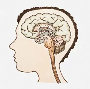
Background imageThe Human Brain Collection: Illustration of human brain

Illustration of human brain

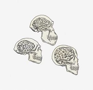
Background imageThe Human Brain Collection: Illustration of skulls of Australopithecus, Homo erectus and Homo sapiens

Illustration of skulls of Australopithecus, Homo erectus and Homo sapiens

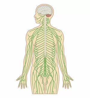
Background imageThe Human Brain Collection: Cross section biomedical illustration of brain and nervous system in adult male

Cross section biomedical illustration of brain and nervous system in adult male

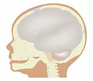
Background imageThe Human Brain Collection: Cross section biomedical illustration of the brain and skull at birth

Cross section biomedical illustration of the brain and skull at birth


All Professionally Made to Order for Quick Shipping


-

Why Choose Us?

We are a leading provider of Art Prints and Photo Gifts since 2003, working in partnership with a range of Sporting Clubs, Charities, Museums and Picture Libraries. A large share of profits from any of their images will go directly towards supporting that charity or club. Our archive of images is carefully curated to bring you a wide range of subjects, including landscapes, wildlife, architecture, and more. We ship from our partner labs in the UK, USA, EU (Netherlands) and Australia.
+

How do I place an order?

Ordering is quick & easy - Just follow 5 Simple Steps:
  1. Find your image: Use our search box or browse our online photo Collections to find the image you want.

  2. Choose your print format: Select your desired product and add it to your cart.

  3. Enter your details: If you're a returning customer, simply enter your email address and password, and we'll fill in your billing and shipping address details. All personal details are held securely and are fully GDPR compliant. As standard, we remove all Personally Identifiable Information after 12 months.

  4. Pay for your purchase: We use state-of-the-art security for online shopping and do not have access to your card details.

  5. Sit back and relax: We'll email you confirmation of your order and when it's dispatched. Registered customers can also track orders in the 'My Account' area.

+

How do I pay for an order?

You can pay for your order with most of the major credit and debit cards, or PayPal. For added security, major financial institutions process payment details separately and securely on our behalf. We do not have access to your online payment card details. Online payments are preferred however we do take phone orders. For UK customers only we accept cheques issued against a UK bank.
+

Is my data safe?

We take data security very seriously. We do not have access to your full card details and all payments are requested over a fully secure connection. Additionally, we fully comply with current European and GDPR legislation, and automatically remove all personal data after at most 12 months (unless you have an account with us and have signed in recently). We also have a strict opt-in policy and would never sell your personal details. Your data is only used to fulfill your order promptly and efficiently.
+

Is wall art ready to hang?

For quick and easy installation all wall art, including framed prints, canvas prints and metal prints are supplied with a ready to hang solution on the back. Generally, saw tooth hangers are applied as they allow wall art to hang flush against the wall. The serrated edge of the hanger prevents the frame from shifting or tilting when hung.
+

Are the photo prints fade resistant?

Yes, we use archival quality photo paper photographic paper for vivid reproduction Prints are an accurate representations of the original artwork, which is preserved for artistic character and authenticity. We guarantee they match previews shown on our web site