Home > Animals > Mammals > Eupleridae > Fossa
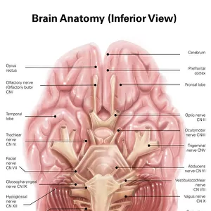
Anatomy of heart interior, frontal section

![]()
Wall Art and Photo Gifts from Stocktrek
Anatomy of heart interior, frontal section
Stocktrek Images specializes in Astronomy, Dinosaurs, Medical, Military Forces, Ocean Life, & Sci-Fi
Media ID 13014471
© Stocktrek Images
Anatomy Aorta Aortic Valve Artery Atrium Biology Biomedical Illustrations Blood Flow Blood Vessels Blue Cardiac Cardiovascular System Chordae Tendinae Circulation Circulatory System Cross Section Cutaway View Cutout Detail Diagram Dissection Endocardium Epicardium Fossa Ovalis Healthcare Healthy Heart Heart Valves Human Anatomy Human Body Parts Human Heart Human Muscles Human Organs Interior Internal Organs Interventricular Septum Left Atrium Left Ventricle Medical Medicine Mitral Valve Muscle Myocardium Organ Papillary Muscles Part Of Pectinate Muscles Physiology Pulmonary Artery Pulmonary Trunk Pulmonary Valve Pulmonary Veins Right Atrium Right Ventricle Semilunar Valve Structure Superior Vena Cava Text Trabeculae Carneae Tricuspid Valve Vein Vena Cava Ventricle Western Script
FEATURES IN THESE COLLECTIONS
> Animals
> Mammals
> Eupleridae
> Fossa
> Posters
> Aircraft Posters
> Cutaway Posters
EDITORS COMMENTS
This print by Stocktrek Images showcases the intricate beauty of the interior of a human heart. The frontal section view provides a detailed glimpse into the complex anatomy and structure of this vital organ, which plays a crucial role in our circulatory system. The image exhibits an array of vibrant colors, with shades of blue representing the veins and red symbolizing arteries. It highlights key components such as the endocardium, myocardium, and epicardium, along with various valves like the pulmonary valve, aortic valve, and mitral valve. Additionally, it features essential structures including ventricles, atria, superior vena cava, pulmonary artery and vein. Every element within this artwork is meticulously depicted to emphasize its significance in maintaining a healthy cardiovascular system. From pectinate muscles to chordae tendinae and trabeculae carneae - each detail contributes to understanding how blood flows through these chambers efficiently. The white background allows for focused observation while emphasizing the intricacy of blood vessels that intertwine throughout this internal masterpiece. This close-up cross-section offers viewers an opportunity to appreciate both the artistic representation and scientific accuracy captured in this photograph. Whether you are studying biology or simply fascinated by human anatomy's wonders, this print serves as an excellent educational tool or aesthetic addition to any healthcare setting or personal collection.
MADE IN AUSTRALIA
Safe Shipping with 30 Day Money Back Guarantee
FREE PERSONALISATION*
We are proud to offer a range of customisation features including Personalised Captions, Color Filters and Picture Zoom Tools
SECURE PAYMENTS
We happily accept a wide range of payment options so you can pay for the things you need in the way that is most convenient for you
* Options may vary by product and licensing agreement. Zoomed Pictures can be adjusted in the Cart.