Fine Art Print > Animals > Mammals > Muridae > Water Mouse
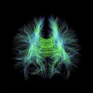
Fine Art Print : Brain fibres, DTI MRI scan C017 / 7035


![]()
Fine Art Prints from Science Photo Library
Brain fibres, DTI MRI scan C017 / 7035
Brain fibres. 3D diffusion tensor imaging (DTI) magnetic resonance imaging (MRI) scan of nerve pathways in the brain. The pathways are highlighted in blue. The brain is seen from the side, with the front of the brain at right. Diffusion tensor imaging measures the direction of water diffusion, which in the brain reveals the orientation of nerve fibres. The technique is also known as tractography, with the resulting image known as a tractogram
Science Photo Library features Science and Medical images including photos and illustrations
Media ID 9340241
© SHERBROOKE CONNECTIVITY IMAGING LAB/SCIENCE PHOTO LIBRARY
Brain Imaging Brain Scan Central Nervous System Cerebral Cerebrum Diffusion Tensor Imaging Dti Scan Fiber Fibers Fibre Fibres Imaging Technique Magnetic Resonance Imaging Mri Scan Mri Scanner Nerve Nerve Fibre Nerves Neural Pathway Neural Tract Paths Pathway Pathways Structural Tractogram Tractography White Matter Brain Neurological Neurology
20"x16" (+3" Border) Fine Art Print
Discover the intricacies of the human brain with our Fine Art Print from Media Storehouse, featuring the captivating image "Brain Fibres, DTI MRI Scan C017 / 7035" by SHERBROOKE CONNECTIVITY IMAGING LAB/SCIENCE PHOTO LIBRARY. This stunning 3D diffusion tensor imaging (DTI) magnetic resonance imaging (MRI) scan brings to life the complex network of nerve pathways in the brain, highlighted in mesmerizing blue. Bring this work of art into your home or office to inspire curiosity and ignite conversations about the wonders of the mind and neuroscience. Each print is produced using high-quality materials and techniques to ensure a vibrant and long-lasting image. Order yours today and embark on a visual journey into the depths of the human brain.
20x16 image printed on 26x22 Fine Art Rag Paper with 3" (76mm) white border. Our Fine Art Prints are printed on 300gsm 100% acid free, PH neutral paper with archival properties. This printing method is used by museums and art collections to exhibit photographs and art reproductions.
Our fine art prints are high-quality prints made using a paper called Photo Rag. This 100% cotton rag fibre paper is known for its exceptional image sharpness, rich colors, and high level of detail, making it a popular choice for professional photographers and artists. Photo rag paper is our clear recommendation for a fine art paper print. If you can afford to spend more on a higher quality paper, then Photo Rag is our clear recommendation for a fine art paper print.
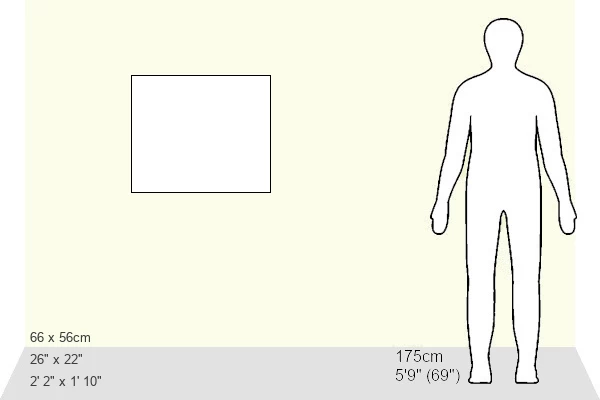
Estimated Image Size (if not cropped) is 50.8cm x 37.5cm (20" x 14.8")
Estimated Product Size is 66cm x 55.9cm (26" x 22")
These are individually made so all sizes are approximate
Artwork printed orientated as per the preview above, with landscape (horizontal) orientation to match the source image.
FEATURES IN THESE COLLECTIONS
> Animals
> Mammals
> Muridae
> Water Mouse
> Posters
> Scientific Posters
EDITORS COMMENTS
This print showcases the intricate network of brain fibres, captured through a cutting-edge imaging technique known as diffusion tensor imaging (DTI) magnetic resonance imaging (MRI). The image, titled "Brain Fibres, DTI MRI Scan C017 / 7035" provides a mesmerizing glimpse into the inner workings of our most complex organ. Highlighted in brilliant blue against a black background, the nerve pathways within the brain are beautifully illuminated. From this side view perspective, with the front of the brain on the right, we can appreciate the astonishing complexity and organization of these neural connections. DTI measures water diffusion direction within the brain to reveal nerve fibre orientation. This innovative technique is also referred to as tractography and produces an image called a tractogram. By visualizing these pathways, scientists gain invaluable insights into how information travels throughout our brains. This extraordinary photograph not only represents a triumph in medical technology but also serves as a testament to human biology and anatomy. It symbolizes health and normalcy while highlighting our ever-expanding knowledge of neurology. Captured by Sherbrooke Connectivity Imaging Lab for Science Photo Library, this image offers us an awe-inspiring glimpse into one of nature's greatest wonders –the human brain– reminding us just how remarkable and intricately interconnected we truly are.
MADE IN AUSTRALIA
Safe Shipping with 30 Day Money Back Guarantee
FREE PERSONALISATION*
We are proud to offer a range of customisation features including Personalised Captions, Color Filters and Picture Zoom Tools
SECURE PAYMENTS
We happily accept a wide range of payment options so you can pay for the things you need in the way that is most convenient for you
* Options may vary by product and licensing agreement. Zoomed Pictures can be adjusted in the Cart.